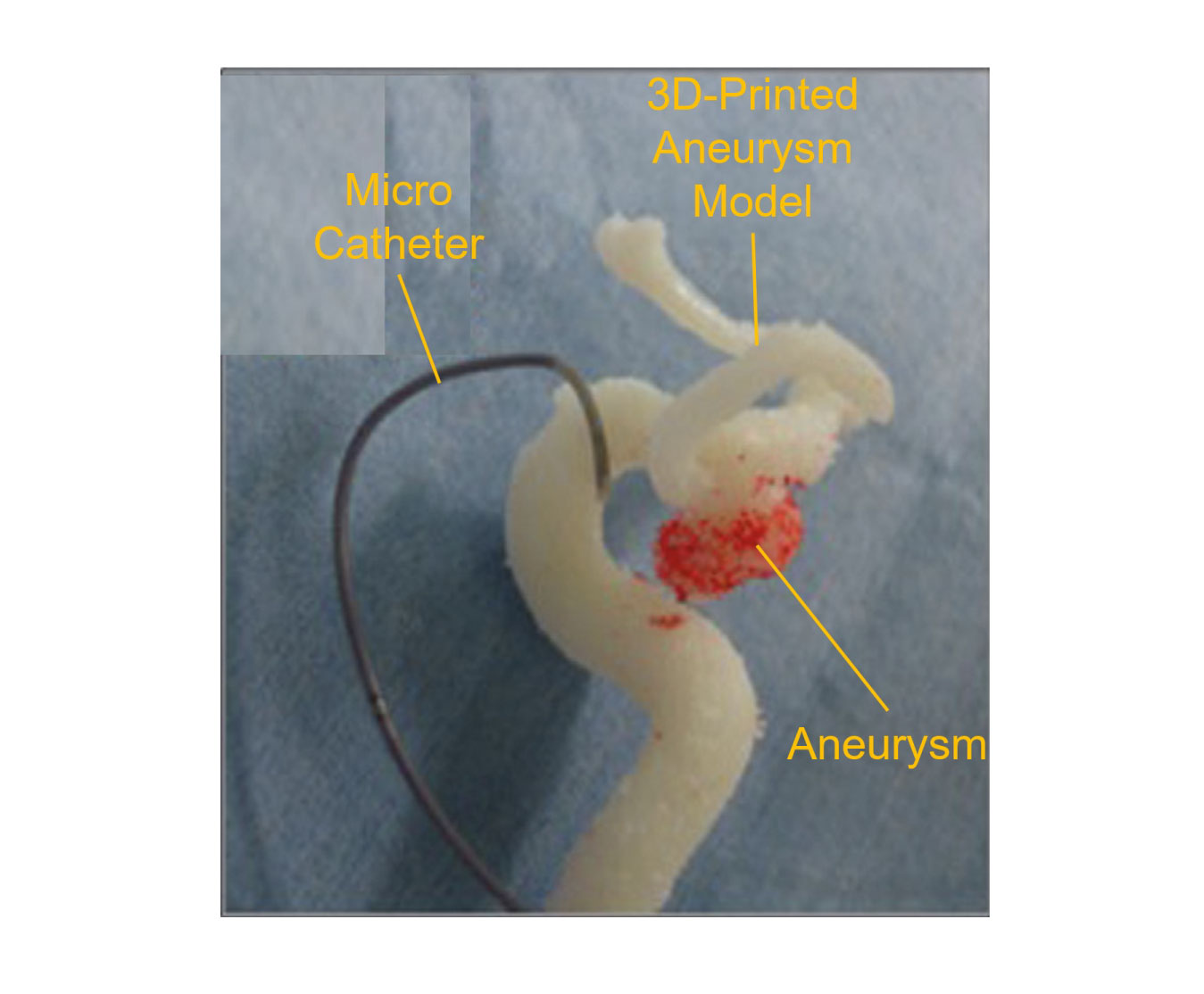
3D-Printed Aneurysm Model
3D aneurysm model is generated from three dimensional medical image data (DICOM data) with its parent arteries using the latest 3D printer. It will be able to understand the aneurysmal morphology or relative position to parent arteries with the actual size. The 3D-printed aneurysm model is valid for the effective micro-catheter shaping in case of endovascular treatment such as coil embolization.
Model Lineup
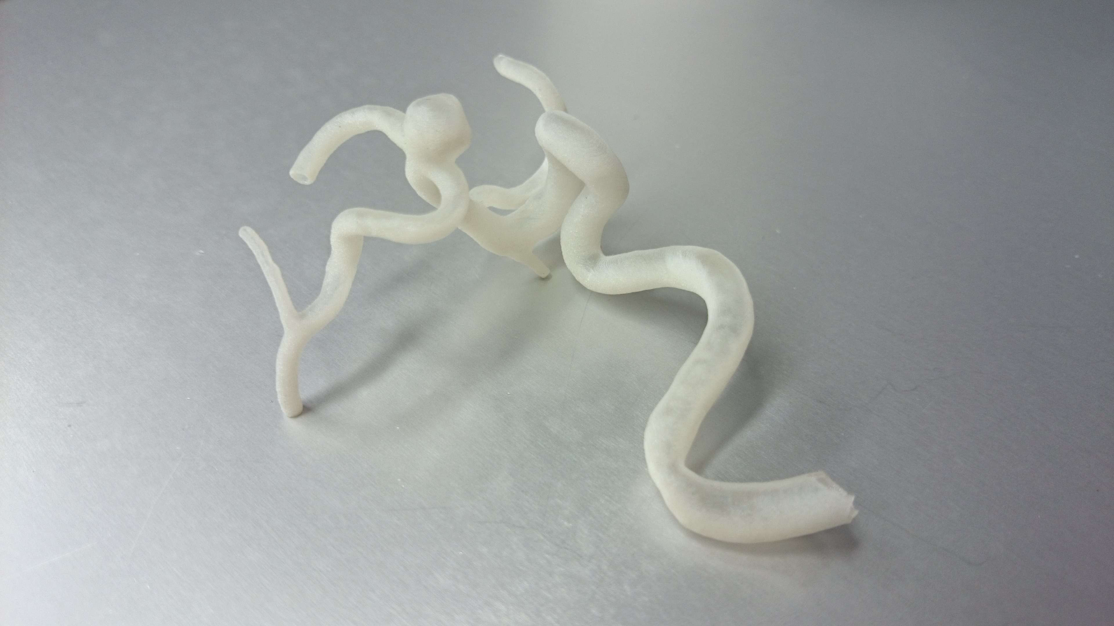
Non-Lumen Model
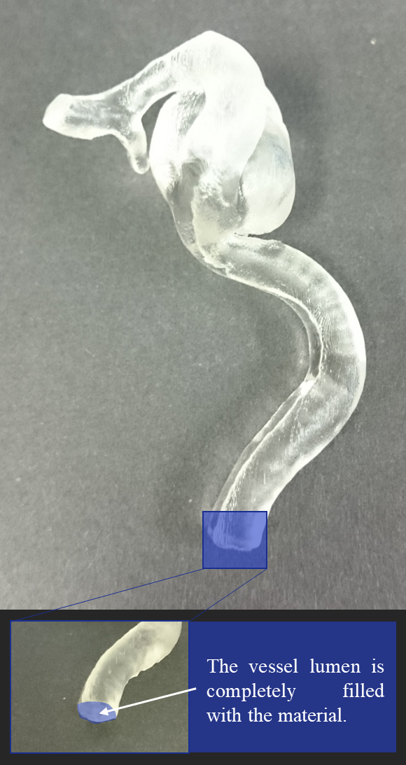
・A simple model with filling the vessel lumen
・Modeled the volume where the contrast medium had flowed
Lumen Model*
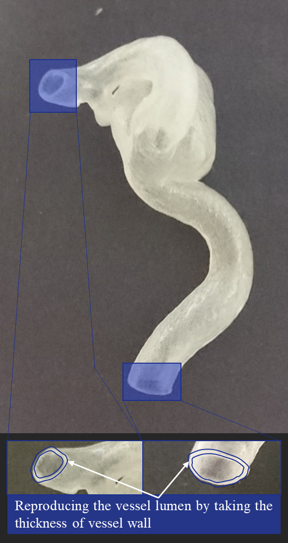
・A model reproducing the vessel lumen
・For checking the fitness of shaped micro-catheter
U-shaped Model**
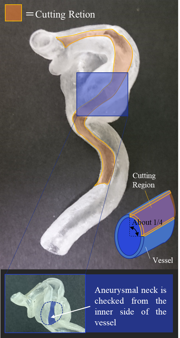
・1/4 cutting model of Lumen Model along the blood vessel
・For checking the fitness of treatment device
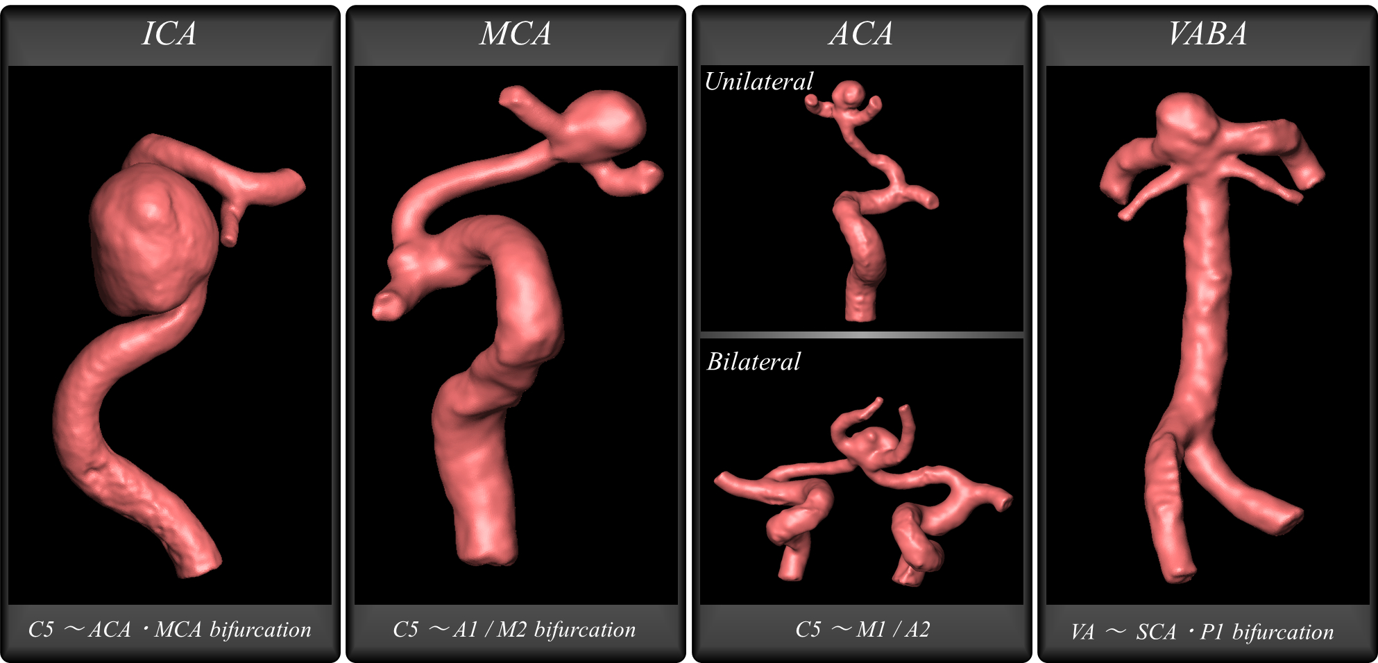
Modeling Region
The modeling region is deffered according to the aneurysm site. The standard regions are as follows:
We will individually correspond for aneurysms in other site.
・MCA: Middle Cerebral Artery … From around C5 to around A1 and M2 bifurcation
・ACA: Anterior Cerebral Artery… From around C5 to around M1 and A2 (Bilateral case is able to modele in single piece)
・VABA: Vartebral Artery and Basilar Artery… From VA to SCA・P1 bifurcation
※We will present the modeling image at the quotation. Please let us know if you want to modify the modeling region.
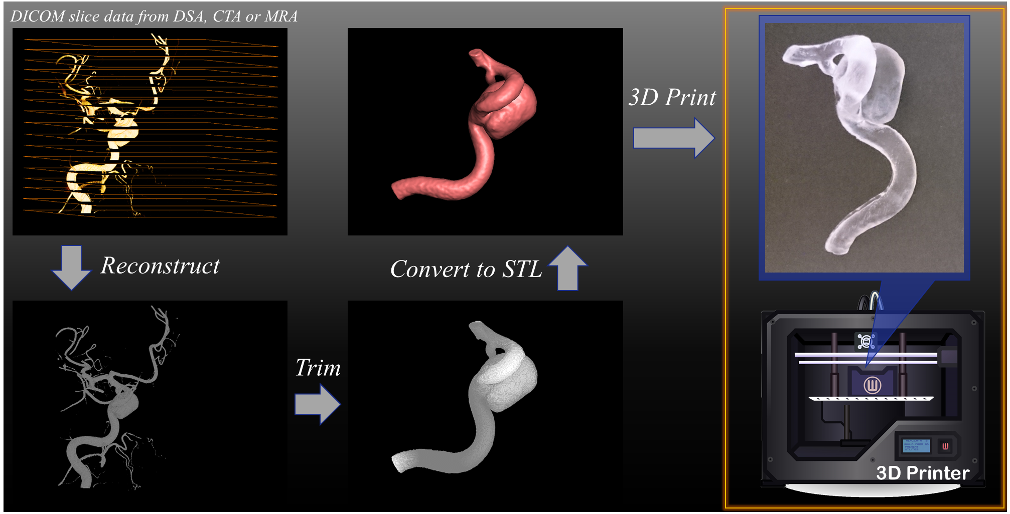
How to Produce ?
The 3D-printed model was generated from the medical imaging data saved in DICOM data, which is obtained from DSA, CTA, or MRA. The aneurysm and vessel geometries are reconstructed from the DICOM slice data. Trimming and smoothing are carried out and the geometry data is converted into STL data format. Using the STL data, 3D-prited aneurysm model is generated by our 3D printer.
How to Use
The catheter position in aneurysm is important to insert the embolic coil into aneurysmal dome at coil embolization. For example, the tip of the catheter is placed at the center of the aneurysm with contacting the parent artery as shown in the figure. The catheter, which is placed such as the figure, is stable and make it easy to insert the coil into aneurysm; the coil will not fall out to the parent artery.
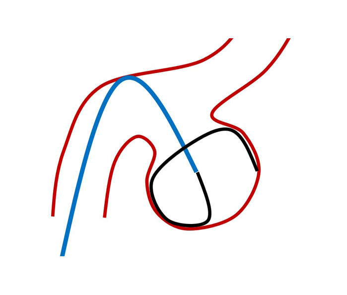
These catheter stability is important to obtain the high packing density aneurysm efficiently. The catheter shaping is carried out to make the catheter place at stable position. The catheter is literally shaped as shown in the next figure.
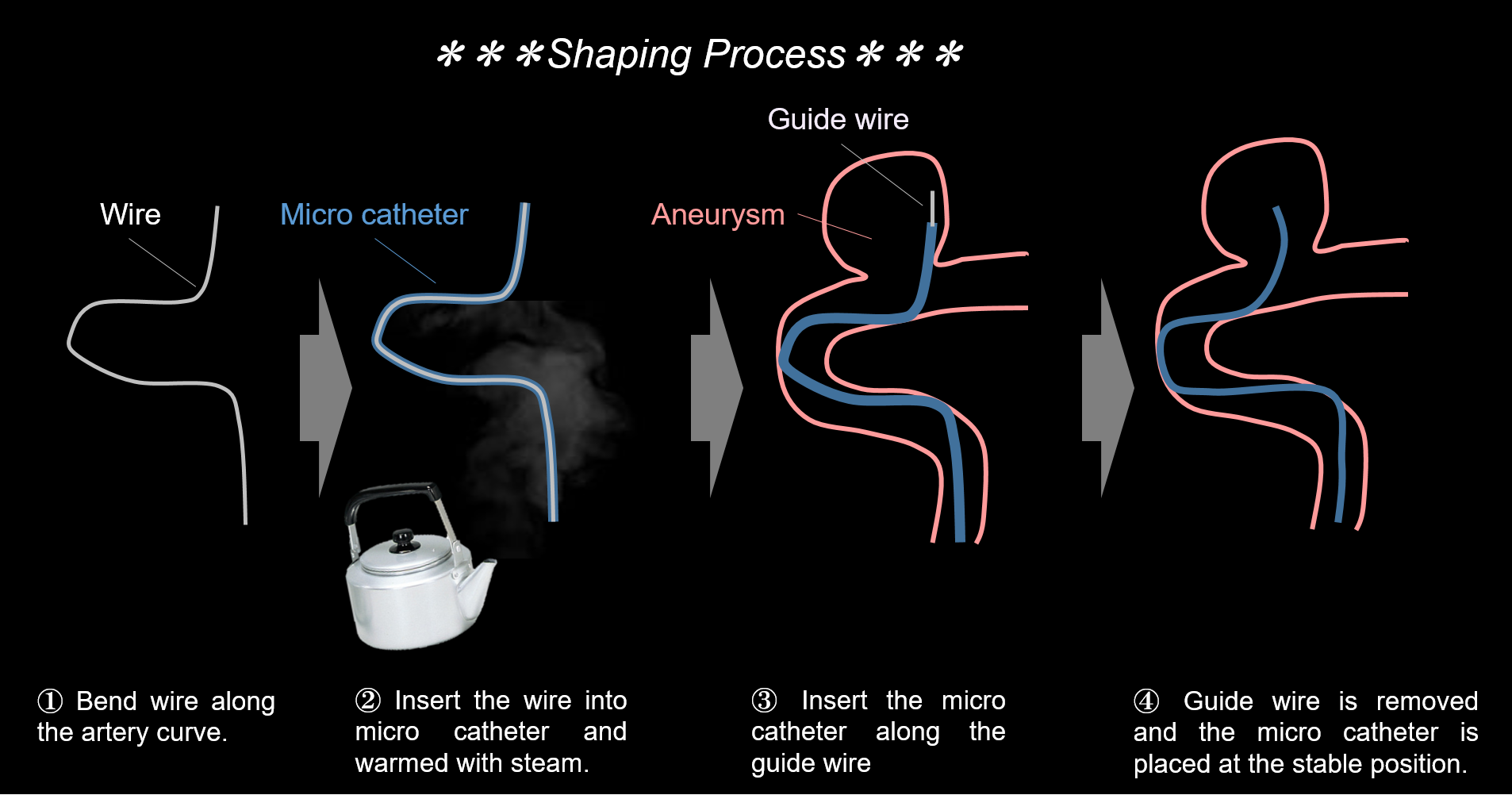
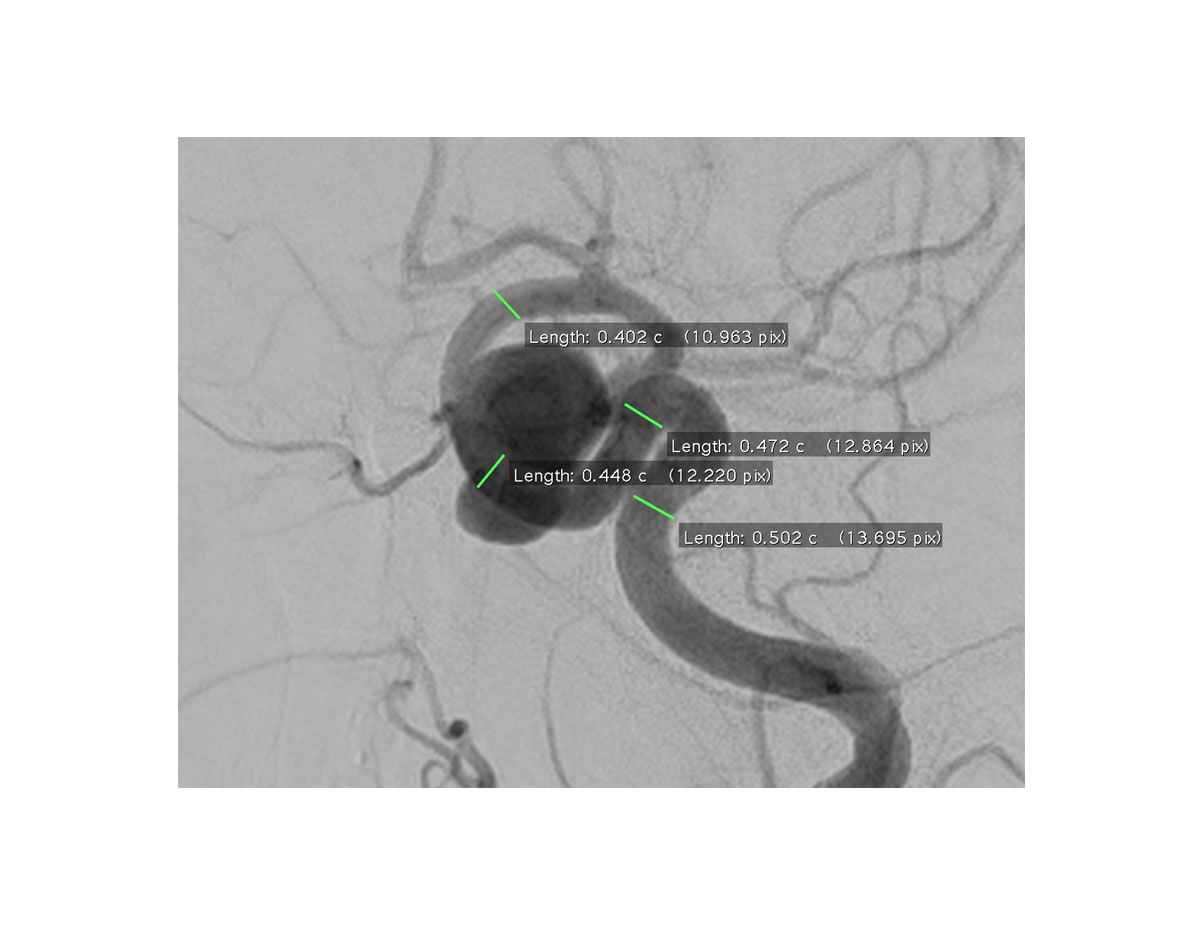
The shaping have been performed referring to the 2D-displayed image of aneurysm like the next figure. However, this method is difficult to comprehend the precise size, depth, and relative position of aneurysm even if the physician is veteran.
The effective and speedy catheter shaping could be carried out using our 3D-printed aneurysm model, which is the same size and geometry with the patient’s real aneurysm. This 3D-printed model is also useful to patient or their families to understand their cerebrovascular disease with their hand and eyes.
Some researchers or doctors reported that the micro catheter shaping using the 3D-printed aneurysm is useful to efficiently perform coil embolization.(Ishibashi T, et al., “Tailor-made shaping of microcatheters using three-dimensional printed vessel models for endovascular coil embolization.”, Comput Biol Med, 2016 Oct 1; 77:59-63.)